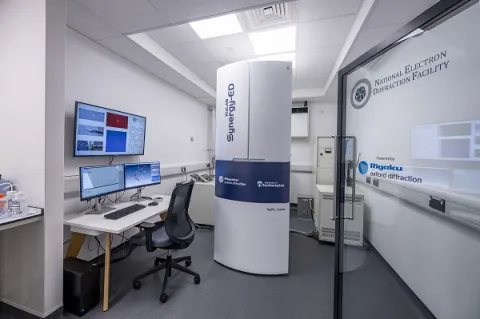
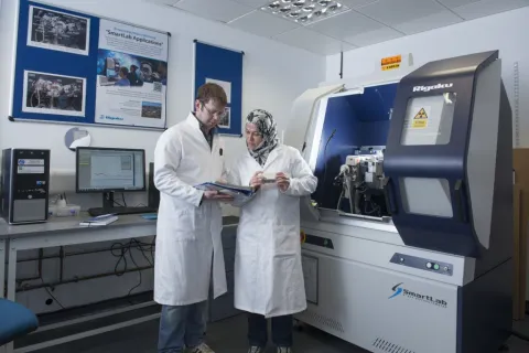
About the X-ray diffraction imaging facility
The X-ray diffraction imaging facility is ideally suited for crystal structure determination, high resolution surface texture mapping, texture pole plotting and rapid sample screening for synchrotron X-ray measurements. The instrument is uniquely configured so that a wide range of measurements can be taken on single crystals, thin films and powdered materials.
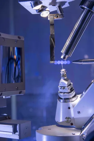
The X-ray diffraction imaging facility consists of a Rigaku MicroMax-007 rotating anode X-ray generator, capable of focusing X-rays down to a spot size of 0.075 millimetre (mm) in diameter. X-rays are imaged with a Dectris Eiger2 area detector, which is capable of single photon counting and can operate at a high frequency of up to 50 Hertz (Hz)
A robotic diffractometer sample mounting stage permits automated and rapid measurement of up to 6 samples in sequence.
Computed tomography (CT) capabilities are provided by the µ-VIS X-ray imaging centre and are unavailable at this facility.