Research
Research groups
Research projects
Completed projects
Researchers:

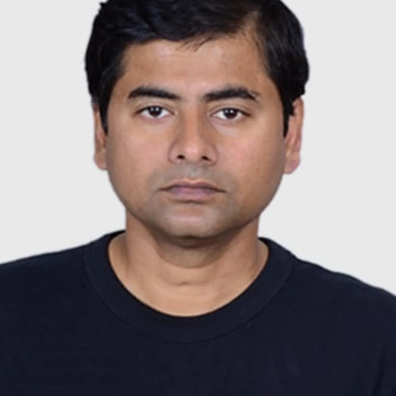
Dr. Dipanjan Bhattacharya has multidisciplinary expertise in the field of microscopy, biophysics, and quantitative image analysis. Presently he is working as a senior research fellow and head of microscopy in the Molecular BioPhotonics & Imaging unit under the Chemical Biology, Diagnostics and Therapeutics research division of the school of chemistry. In 2021, Dr. Bhattacharya is a co-PI in the EU funded FET Open grant under European Union’s Horizon-2020, for the OrganVision project along with his collaborators from four different European countries. His position at the University of Southampton is funded by the EPSRC funded Transformative Imaging for Quantitative Biology (TIQBio) Partnership project.
OrganVision aims to develop a technological platform for real-time visualizing and modelling of fundamental process in living organoids towards new insights into organ-specific health, disease, and recovery. At the University of Southampton, Dr Bhattacharya is leading the WP4 and is responsible for developing ‘OrganLab’ for the OrganVision project. He is developing microscopy system for fast label free imaging and integrating biocompatible chambers for live engineered heart tissue (EHT) imaging. The OrganVision project is a consortium of researchers from the Arctic University of Tromso (UiT) - Norway (Coordinator), the University of Southampton- the United Kingdom, the University of Barcelona - Spain, The University Medical Center Hamburg-Eppendorf (UKE) - Germany, The University hospital of North Norway (UNN) - Norway, a small and medium enterprise (SME) from Italy - 3rd Place and a high-tech Spinoff company from Germany - JenLab. This consortium consists of researchers from diverse disciplines – physicists, bioengineers, , biophysicists, medical doctors, computational scientists etc.
The Transformative Imaging for Quantitative Biology (TIQBio) is an EPSRC funded new Prosperity Partnership between the University of Southampton, a global pharmaceutical company - AstraZeneca and a technology company M Squared Life, to revolutionise the imaging technology to accelerate drug discovery. This project aims to develop tools to provide live, high-resolution 3D images on a large scale to determine the impact of new drug candidates and their effectiveness in treating various conditions. Dr. Bhattacharya is involved in developing super resolution light sheet microscopy for both fluorescent and label-free imaging modalities towards high throughput imaging. Towards this he is developing both theoretical and microscopic framework of resolution enhancement, aberration correction, better sample penetration and optical beam shaping. He is also involved in developing deep-learning based approaches for aberration correction of the optical imaging system as well as for the applications related metabolic profiling.
Along with these, he is also involved in developing innovative image analysis technologies for different biological and biomedical applications towards disease diagnosis and cure.
Professional background:
Dr. Bhattacharya did his master’s in physics at the University of Kalyani, India. He then joined Raman Research Institute, Bangalore for his Ph.D. in Biophysics to pursue a collaborative project with National Center for Biological Sciences, TIFR, Bangalore. His Ph. D thesis was aimed at understanding the role of histone dynamics and its functional implementation in live cells. During his Ph. D, he developed diverse microscopic techniques based on both force and fluorescence microscopy for quantitative biological applications.
For his postdoctoral work, he joined Prof. Paul Matsudaira’s lab at the Massachusetts Institute of Technology (MIT) in the Bio-Engineering dept. and worked in Whitehead Bioimaging Center. During this period, he enhanced his skills in Computational Biology and high-end Image Processing. Following this, he moved to Singapore and joined Singapore-MIT Alliance for Research and Technology Center (SMART), a newly developed campus of MIT in Singapore. At SMART he worked with Prof. Paul Matsudaira, Prof. George Barbastathis and Prof. Peter So, for developing diverse microscopic techniques, like Light-sheet microscopy (DSLM) with structured illumination, Volume Holographic Microscopy (MVH system), Confocal Phase Microscopy, Image Cytometry system, etc for various biomedical applications. He used these homebuilt imaging systems to understand the mechano-biological cues of the convergence extension process in Zebrafish embryogenesis. Simultaneously, he also developed numerous computational tools for quantitative automated analysis of electron microscopy images. From 2012-17, he was jointly appointed as a research scientist in Mechanobiology Institute, Singapore and Singapore-MIT Alliance for Research and Technology Center (SMART). From 2018-2022, he joined IFOM center (IFOM ETS - the AIRC Institute of Molecular Oncology) at Milan to lead the Imaging development unit. At IFOM he was developing innovative imaging and image analysis tools for cancer biology and other biomedical applications. From January 2023, he has moved to University of Southampton as a senior research fellow and the head of microscopy in the Molecular BioPhotonics & Imaging unit under the Chemical Biology, Diagnostics and Therapeutics research unit.