
Histology microscopes

More Information

About the Histology microscopes

The facility provides histological microscopes for colourimetric imaging. We also have additional equipment for the sectioning, preservation and processing of specimens. 
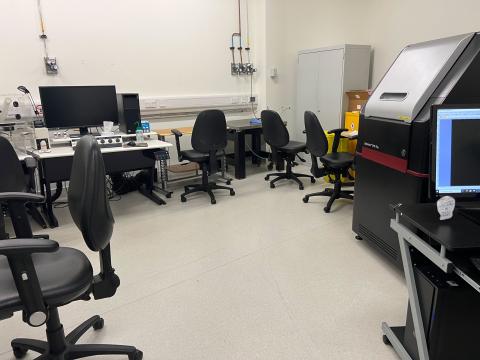
Preparation room

Our prep room is equipped with: 

  • tissue culture equipment, 
  • specimen storage solutions 
  • histology prep equipment

Part of: Imaging and microscopy centre

Technical specification

Nikon E400

Modality: Brightfield Transmitted light

Modification, numerical aperture and immersion

  • 4x, 0.13, air
  • 10x, 0.3, air
  • 20x, 0.5, air
  • 40x, 0.75, air

Detector: QImaging Retiga 2000R CCD colour camera 1600 x 1200 pixels.
 

Specimen format: Standard glass slide
 

Leica CM 1860 UV cryostat

  • section thickness - 1-100 µm (micrometres)
  • total specimen feed - 25 mm (milimetres)
  • vertical stroke
  • max specimen size - 55 x 55 mm
  • UV disinfection - yes
  • temperature range - 0 to -35°C

Leica 3050S Cryostat

  • temperature setting range: 0°C to -40°C
  • maximum specimen size: 40x 55 mm
  • section thickness setting: 0.5μm to 300μm 
  • trimming: 5 to 150 μm ± 0,5 μm in steps of 5, 10, 30, 50, 100 and 150 μm
  • motorized coarse feed at two speeds: 500 μm/s and 1,000 μm/s
  • horizontal specimen feed: 25 mm
  • vertical specimen stroke: 59 mm
  • specimen retraction: 50 pm
  • specimen precision orientation by 8° (x/y/z axis)
  • UV disinfection – No

Cutting speed and range

0.1 mm/s to 170 mm/s, 0.1 mm/s to 100 mm/s, Vmax: 210 mm/s

Leica TP1020 Tissue Processor

The Leica TP1020 is an automatic tissue processor. It is used for the fixation, dehydration and infiltration of histological tissue samples with fixatives, alcohol, solvents and paraffin wax. The processor is made up of 12 reagent stations that are used to hold various concentrations of solvents, HistoClear and paraffin wax. Stations 11 and 12 are heated, temperature-controlled wax baths that can be filled with either wax pellets or molten paraffin wax.

  • wax baths – 2 (3 optional)
  • wax baths capacity – 1.8 litres
  • wax baths temperature range - 45°C to 65°C
  • standard tissue basket capacity – Max 100 cassettes
  • excess temperature cut-out - 75 °C ± 4 °C
  • reagent containers – 10 (9)
  • reagent container capacity – 1.8 litres
  • standard tissue basket  - 1 (2 optional)
  • number of programs – 9, freely selectable
  • programmable infiltration time per station – 99h 59 min
  • delayed start – 9 days
  • drain time – 60 s

Leica RM2235 Manual Rotary Microtome

  • section thickness setting: 1.0 – 60 µm
  • 1 to 10 µm in 1 µm increments
  • 10 to 20 µm in 2 µm increments
  • 20 to 60 µm in 5 µm increments
  • specimen retraction: approx. 40 µm, can be deactivated
  • vertical stroke: 70 mm
  • trim settings: 10 µm, 30 µm
  • horizontal specimen feed: approx. 30 mm / 1.18 inches
  • vertical stroke: 70 mm / 2.76 inches
  • maximum specimen size: 45 x 50 mm / 1.77 x 1.97 inches

Leica EG1150 H Paraffin Embedding Station

The Leica EG1150 H is a modern paraffin embedding station with microprocessor control system. It is designed for embedding histological tissue specimens in molten paraffin for use in pathology laboratories and only for the following tasks: 

  • melt solid paraffin for sample embedding and maintain the molten paraffin at the required temperature. 
  • pour paraffin into embedding moulds where the specimens are placed. 
  • heat and maintain the temperatures of embedding cassettes with specimens and moulds as well as the required forceps.
  • paraffin reservoir – approx. 3L
  • cassette tray – approx. 100 cassettes
  • embedding mould tray – approx. 50 embedding moulds

Contact us

Dr Mark Willett - Highfield
Georgie Dawes - Southampton General Hospital

Highfield

Building 85, Highfield Campus, Southampton SO17 1BJ
We’re open Monday to Friday 09:00 to 17:00 UK time.

Southampton General Hospital

Back
to top